
If your jaw lacks enough bone, a dental implant can fail or sit poorly. A bone graft rebuilds lost jawbone so an implant has a strong, stable foundation — you need one when imaging shows insufficient bone height, width, or density.
You’ll learn what grafting looks like, the common reasons you might need it, and how it affects timing, recovery, and long-term implant success. This article will help you decide whether grafting is likely for your situation and what to expect next.
Essential Role of Bone in Dental Implant Success
Strong, healthy jawbone gives your implant a stable base and helps the metal post fuse with bone. If your bone is thin, soft, or low in height, your implant may not seat firmly or last long.
How Bone Quality Impacts Implant Stability
Bone quality affects how well an implant integrates and bears chewing forces. Dense, thick bone lets the implant lock in immediately and reduces micromovement during healing — both key factors your provider of dental implants in Trinity, FL will assess before recommending a treatment plan. That lowers the risk of failure and speeds up the time before the crown can be placed.
Poor bone density or thin cortical bone increases the chance that the implant will loosen or tilt. You may feel pain, or the implant may fail months after placement. Your dentist checks density with X-rays or a CBCT scan to measure both height and thickness.
If bone is marginal, your clinician may use wider or longer implants, change implant position, or recommend a bone graft to add volume and create a stronger foundation.
Indicators of Insufficient Jawbone for Implants
You may need a bone graft when imaging shows low ridge height, reduced bone width, or large areas of resorption after tooth loss. Common signs include shallow ridges where the crest feels narrow under the gums and X-rays that show thin bone around tooth roots or sinus proximity in the upper jaw.
Other indicators include advanced periodontal disease, long-term tooth loss that caused bone shrinkage, and trauma that removed bone. Your dentist also looks for vertical defects near neighboring teeth and insufficient bone to fully cover an implant thread.
Clinical tests include probing for ridge width, measuring vertical height on scans, and assessing sinus or nerve location. If these measures fall below implant requirements, grafting or alternative techniques will be recommended.
Understanding Bone Graft Procedures
A bone graft builds jawbone where it is weak or missing so an implant can sit firmly. You will learn what graft materials are used, how the surgeon places them, and how long healing usually takes.
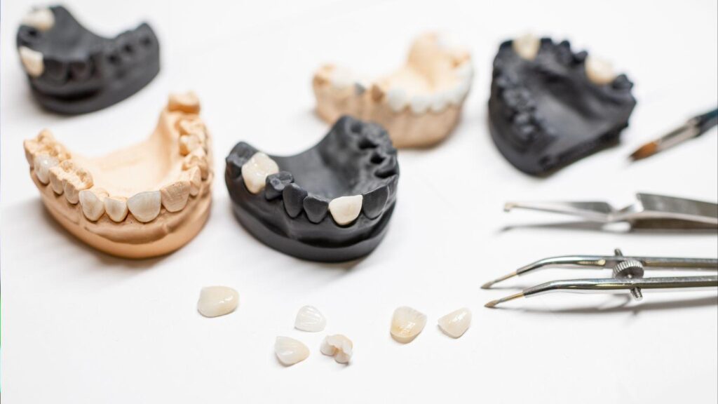
Types of Bone Grafting Materials
You may get one of four main graft materials:
- Autograft (your own bone): Often from the chin, lower jaw, or hip. It has the best chance to fuse because it contains living cells and growth factors.
- Allograft (donor human bone): Taken from a tissue bank. It avoids a second surgical site and still supports new bone growth.
- Xenograft (animal bone): Usually from bovine sources. It provides a durable scaffold that your bone gradually replaces.
- Alloplast (synthetic materials): Made of calcium phosphates or other compounds. They reduce infection risk and are widely available.
Your dentist will choose based on how much bone you need, the site of the graft, your health, and whether you can tolerate a second surgical site. Costs, healing speed, and fusion reliability vary by material.
Techniques Used in Dental Bone Grafting
Surgeons use several common techniques:
- Socket preservation: Placed right after a tooth extraction to keep the ridge from collapsing.
- Ridge augmentation: Adds width or height to the jaw where bone has resorbed.
- Sinus lift (sinus augmentation): Lifts the sinus membrane and places graft material in the upper back jaw to create height for implants.
- Block grafts: A solid piece of bone (often autograft) is fixed to the jaw with screws when large volume is needed.
Procedures can be done under local anesthesia, IV sedation, or general anesthesia depending on complexity. Your surgeon will suture the site and may use membrane barriers to protect the graft and guide bone growth. Expect imaging (X-rays or CBCT) before and after to track graft integration.
Typical Timeline for Recovery
Initial recovery takes about 1–2 weeks. You will likely have swelling and mild pain that peaks in 48–72 hours and then improves with medication and cold packs.
Stitches may be removed or dissolve within 7–14 days.
Bone healing and remodeling take longer:
- 3 months: Early bone formation begins; some implants can be placed if graft was small.
- 4–9 months: Most grafts integrate and reach strength for implant placement.
- 9–12+ months: Full maturation for large grafts or when the graft source was less active.
Follow-up visits and imaging confirm integration before implant surgery. Avoid smoking and follow your surgeon’s diet and oral care instructions to help healing.
Factors That Necessitate Bone Grafting
You may need a bone graft when the jaw lacks enough height, width, or density to hold an implant. Common causes include bone loss after extraction, long-term gum disease, or injuries that damage the bone structure.
Bone Loss From Tooth Extraction
When a tooth is removed, the socket and surrounding bone start to shrink within weeks to months. If you wait months or years to replace the tooth, the ridge can narrow and lose height, leaving too little bone to stabilize an implant screw.
Dentists often measure the ridge with X-rays or a CBCT scan to see how much bone remains. If the socket has collapsed or the ridge is thin, your surgeon may place graft material to rebuild height and width. This creates a stronger foundation and improves implant positioning and long-term stability.
You might get a graft placed at the same time as the extraction (socket preservation) or later as a separate procedure. Healing typically takes several months before an implant can be placed.
Periodontal Disease and Jawbone Deterioration
Advanced gum disease destroys the bone that supports teeth. If you have chronic periodontitis, pockets form around teeth and bacteria eat away bone over time. This bone loss can be local (around a few teeth) or widespread.
Your dentist will assess bone loss with probing and imaging. If disease removed significant bone where you want an implant, grafting rebuilds the lost volume and addresses uneven bone contours. Treating the infection first is essential; grafts placed into infected sites have higher failure risk.
Grafts for periodontal defects often use bone chips, membranes, or growth factors to encourage new bone. The goal is to restore enough bone to keep an implant stable and to create healthy gum support.
Effects of Trauma or Injury on Bone Structure
A facial injury or tooth fracture can crack, displace, or remove sections of jawbone. If you had a fractured alveolar ridge, an avulsed tooth, or reconstructive surgery, the remaining bone may be irregular or insufficient for an implant.
You will need imaging to map the defect and plan the graft. Trauma-related grafting can require shaping the jaw, adding volume, or correcting angled bone to allow correct implant placement. In some cases, block grafts (solid bone pieces) are used instead of particulate grafts to rebuild larger defects.
Healing time after trauma grafting varies with defect size and graft type. Your surgeon will schedule follow-up scans to confirm bone integration before placing the implant.
Considerations and Outcomes for Patients
You will weigh risks, benefits, and the care needed after the procedure. Knowing what can go wrong, what good bone support delivers, and how to care for the site helps you make better choices and recover more predictably.
Risks and Complications
Bone grafting carries common surgical risks: infection, bleeding, swelling, and pain. These typically peak in the first 48–72 hours and respond to antibiotics, ice, and pain medicine your clinician prescribes.
Graft-specific problems include graft failure (lack of new bone forming) and partial resorption (some graft material shrinking). Smoking, uncontrolled diabetes, and poor oral hygiene raise these risks. Tell your dentist about medications like blood thinners or immune suppressants, as they affect bleeding and healing.
Less common but important risks are nerve injury (numbness or tingling) and sinus perforation for upper jaw grafts. Your surgeon will assess anatomy with imaging to lower those chances. Report increasing pain, fever, or unusual drainage right away.
Expected Benefits of Adequate Bone Support
A successful graft creates enough height and width of jawbone to hold an implant securely. This stability lowers the chance of implant loosening and improves chewing function. You gain a stronger foundation for a crown that looks natural and fits well with nearby teeth.
Good bone also helps long-term oral health by supporting surrounding gums and preventing further bone loss. In many cases, proper grafting lets you avoid removable dentures and preserves facial shape. Success rates are high when you follow pre-op and post-op instructions and manage health issues like diabetes.
Post-Surgical Care Recommendations
Follow your clinician’s written instructions for the best outcome. Generally, you should avoid brushing the surgical site for a few days, rinse gently with saltwater or a prescribed mouthwash, and use ice packs for 24 hours to reduce swelling. Eat soft foods and avoid chewing on the grafted area until cleared.
Take all prescribed antibiotics and pain medicine exactly as directed. Do not smoke for at least several weeks; nicotine cuts blood flow and reduces healing. Attend follow-up visits for suture removal and imaging so the team can confirm bone is forming and decide when to place the implant.